


Wisdom Tooth Extraction,
It is planned according to the location of the tooth and the risk it poses.

It examines the eruption of the wisdom tooth, its relationship with the neighboring teeth and the nerve canal in the lower jaw. We make the follow-up or extraction decision based on clinical findings.

20-year-old dental examination
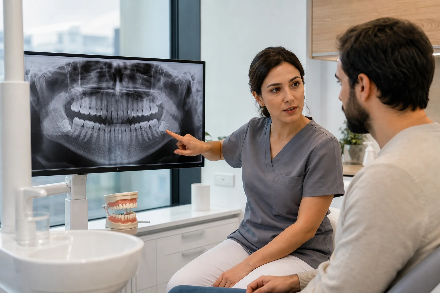
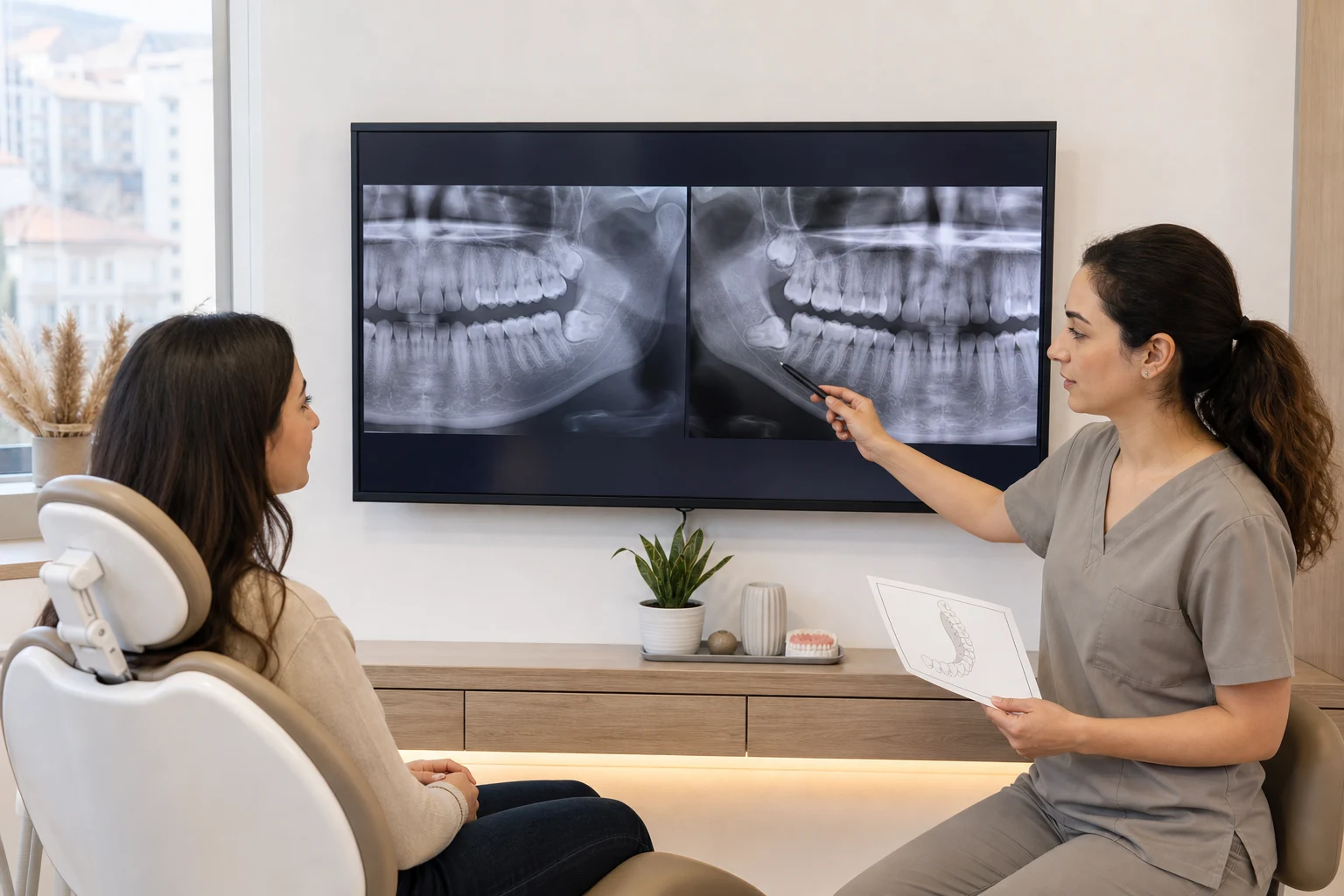
Dentist explaining impacted lower wisdom tooth to patient on panoramic x-ray
Before the decision to shoot

We examine together the angle of the tooth, the surrounding bone, the adjacent molar and its relationship with anatomical structures.

Wisdom tooth evaluation

In wisdom teeth
How is the decision to follow or shoot made?

Wisdom teeth are located at the very back of the jaw. When there is not enough space for eruption, the tooth may remain completely within the bone, partially erupt, or be inclined towards the adjacent tooth.

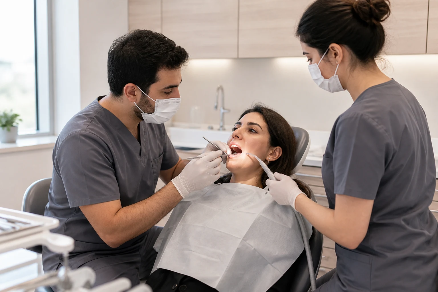
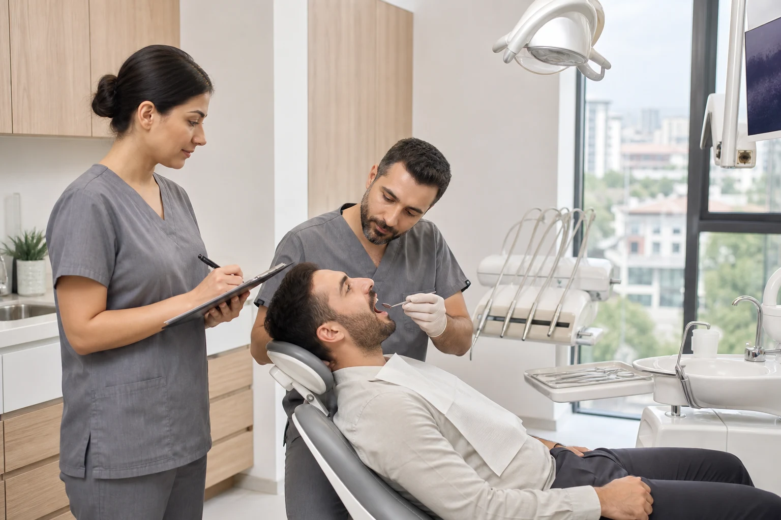
The team that clinically examined the lower wisdom teeth area
The gingiva on the semi-erupted tooth, cleanability, mouth opening and sensitivity of the surrounding tissues are evaluated during the clinical examination.

Impaction alone is not always a reason for extraction; signs of disease or damage are important in the decision.

Pain and swelling, recurrent gum infection, food accumulation that cannot be cleaned, decay in the wisdom tooth or the adjacent tooth, and pathological changes around the tooth are investigated.

In the lower 20's teeth, the roots are connected to the nerve canal; In the upper wisdom teeth, their relationship with the surrounding anatomical structures is examined through images. For teeth that do not cause problems, regular follow-up options can be considered.

A single history of inflammation does not always mean surgery.The severity of the complaint, whether it recurs or not, and its effect on surrounding tissues are considered together.

01
Riding statusFully erupted, half-buried or buried
02
neighbor toothRot, root damage and cleanability
03
anatomical relationshipNerve canal, bone and surrounding tissues
04
clinical findingHistory of pain, infection, swelling and recurrence
Surgical process

Wisdom tooth extraction process
proceeds with imaging and surgical planning.

Standard extraction and impacted tooth surgery are not the same procedure. The eruption level of the tooth, its root shape, its angle within the bone and its relationship with anatomical structures determine the method.

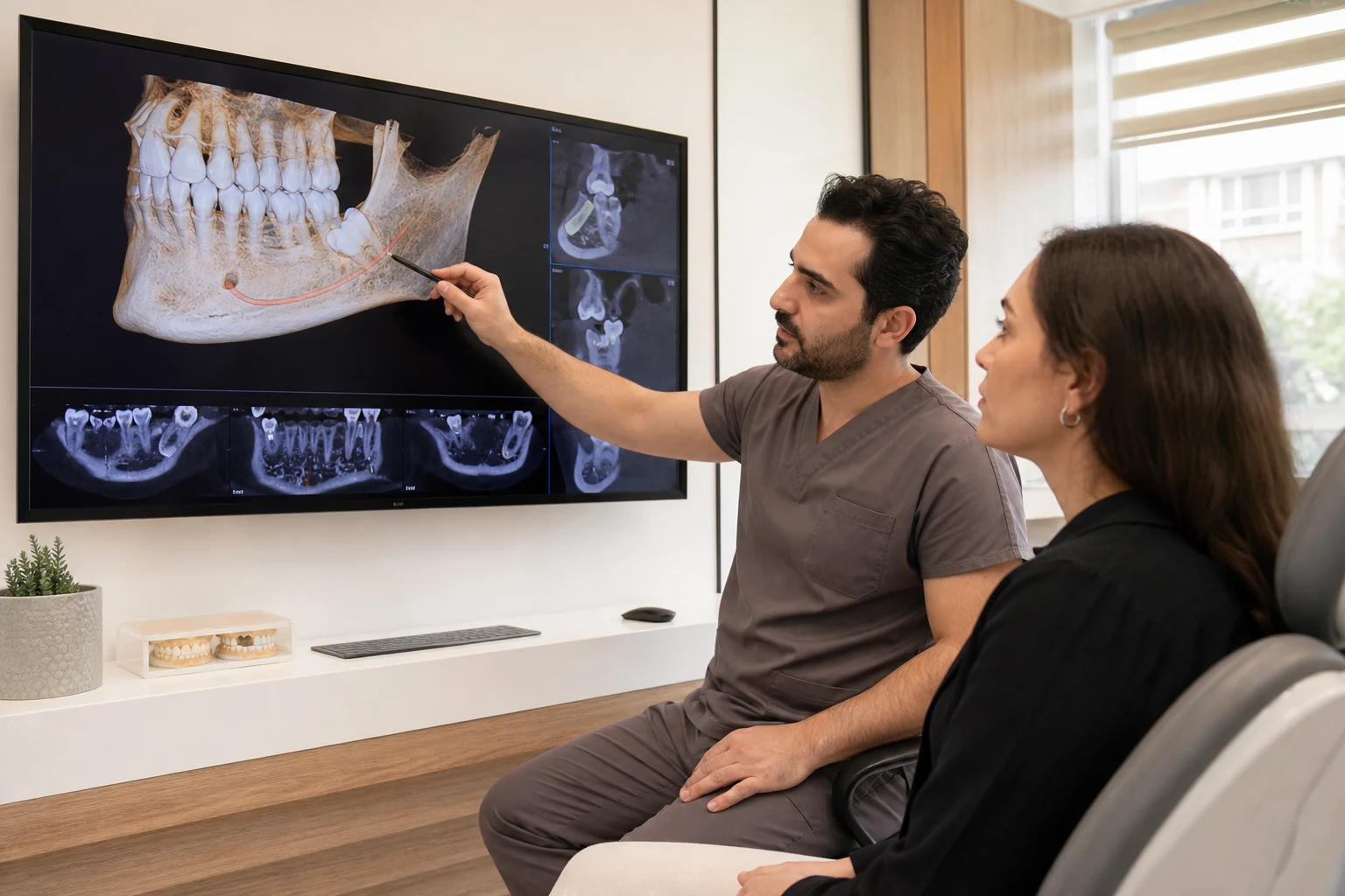
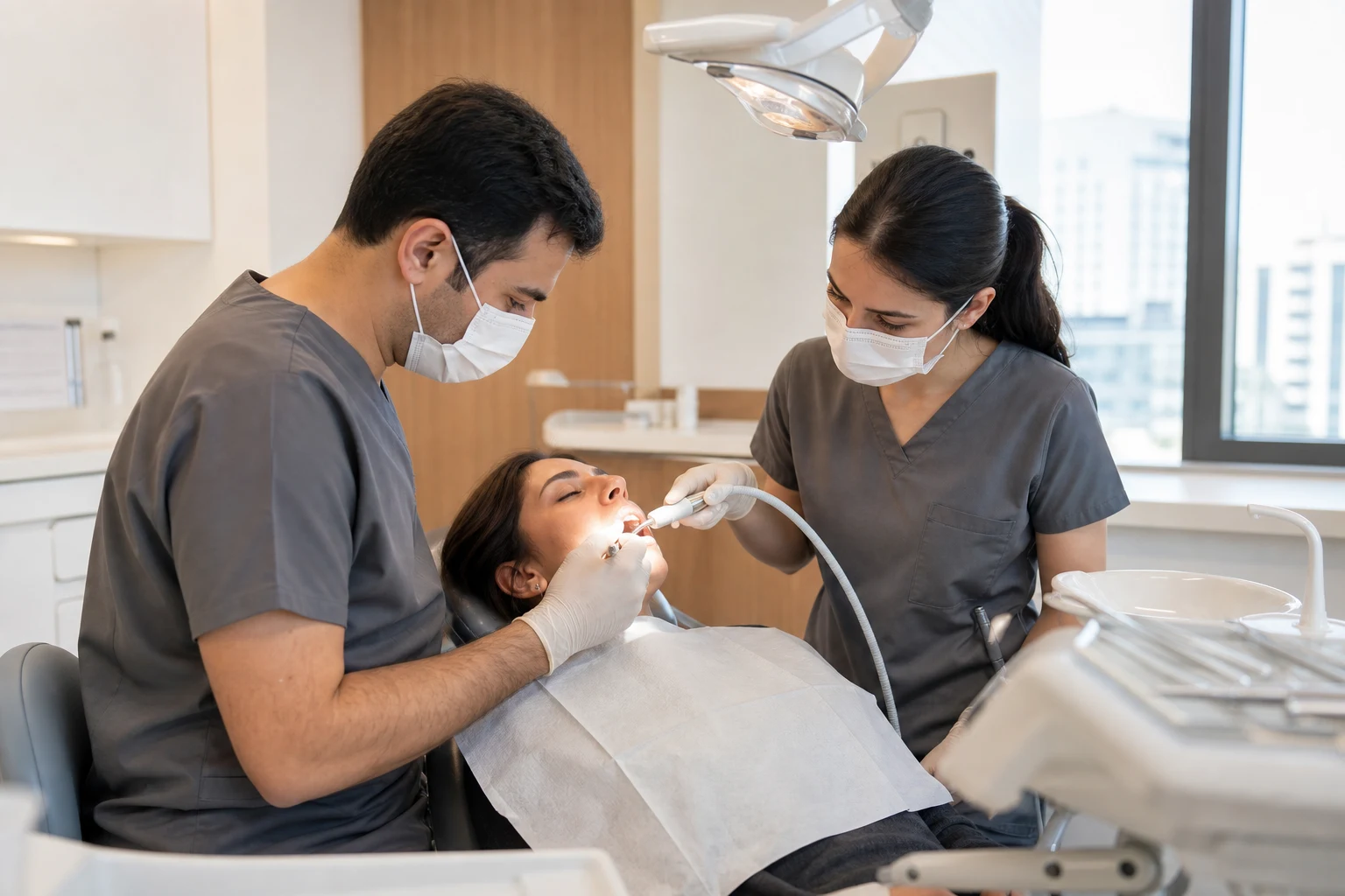
Patient explaining his complaint about wisdom teeth in his lower jaw Dentist evaluating the relationship of the impacted wisdom tooth with the nerve canal in a three-dimensional image The team that performs local anesthesia and sterile preparation before wisdom tooth extraction Clinical team performing wisdom tooth extraction in a controlled manner
01 / Complaint and historyWhen did the symptoms begin?
and we listen to see if it repeats.
01 — 04
  1. 01
    inspection

    Clinical evaluation

    Listens to the history of pain, swelling, and infection. We clinically examine the eruption area, gums, adjacent teeth and mouth opening.

  2. 02
    Panoramic and 3D recording when necessary

    Surgical mapping

    He examines the panoramic x-ray. We can plan three-dimensional imaging in close nerve relationship where two-dimensional imaging is insufficient.

  3. 03
    Secure procedure plan

    Local anesthesia and preparation

    We re-check the health history, anesthetize the procedure area and prepare a sterile surgical procedure appropriate to the eruption level of the tooth.

  4. 04
    Standard or surgical extraction

    Surgical removal of the tooth

    Provides controlled access to gum and bone when necessary. We remove the tooth by dividing it into one piece or sections and close the area.

Tracking or extraction?

Impacted wisdom teeth,
unbearable in any case.

A wisdom tooth with no signs of disease does not need to be routinely extracted. The decision for follow-up or surgery is personalized with current examination and imaging findings.

Dentist monitoring a problem-free impacted wisdom tooth with comparative images
01 / Tracking option

If there is no sign of disease
controlled monitoring

Some impacted teeth that do not cause pain, infection, or damage to surrounding tissues can be left in place. The follow-up decision is evaluated together with the cleanability of the tooth and its future risks.

clinical review
Evaluation of gums, oral hygiene and new complaints
Image comparison
Monitoring whether there are changes in the tooth and surrounding structures
Update of the decision
Reconsidering options if a new disease finding occurs
02 / Extraction and review

Surgery when necessary,
followed by planned follow-up

Extraction may be evaluated when recurrent infection, untreatable decay, damage to the adjacent tooth, or pathological change around the tooth is detected. Post-procedure recommendations are personalized based on the extent of surgery.

first recovery
Clot protection, procedure of nutritional and hygiene recommendations
Expected complaints
Monitoring temporary pain, swelling and limitation of jaw movement
Cases requiring review
Increasing pain or swelling, fever, bad taste, and bleeding that does not stop
Clinical team checking recovery after wisdom tooth extraction

Frequently asked questions

Wisdom teeth:
Curiosities.

Follow-up, imaging and extraction decision. It is personalized after evaluating the position of the tooth, the findings it produces and the surrounding anatomical structures.

No. Some impacted wisdom teeth that show no signs of disease or damage can be monitored clinically and radiographically. The extraction decision considers pain, recurrent infection, decay, damage to the neighbouring tooth, cyst-like changes and other clinical findings.

Recurrent gum infection, pain and swelling, food accumulation that cannot be cleaned in the area, decay in the wisdom tooth or the adjacent tooth, root damage or pathological changes around the tooth are situations in which extraction may be evaluated. The final decision is made after examination and appropriate imaging.

No. Clinical examination and panoramic x-ray may be sufficient for most evaluations. If the roots of the lower wisdom teeth appear close to the nerve canal or if the two-dimensional image does not provide sufficient information for the surgical plan, three-dimensional imaging may be requested.

The procedure is usually performed under local anesthesia. If the tooth is completely erupted, standard extraction can be applied. If it is buried under the gum or bone, controlled access can be provided to the area and the tooth can be disassembled and removed when necessary. The scope of the procedure varies depending on the location of the tooth.

Depending on the extent of the procedure, temporary pain, swelling, jaw stiffness and difficulty chewing may occur. The course of these complaints varies from person to person. In cases of increasing pain or swelling, fever, bad taste or persistent bleeding, the clinic should be contacted without delay.

The roots of the lower wisdom teeth may be close to the nerve canal that carries the sensation of the lip and jaw tip. This relationship is evaluated on images and personal risk is explained before the procedure. When a close relationship is detected, the surgical approach and alternatives are planned in detail.